CPC Case III
Provided By: Dr. Ray Melrose - 2011-05-18
Clinical History
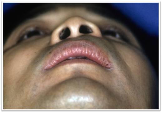
15 year old Hispanic boy
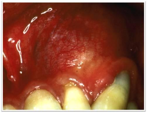
Painless left labial swelling noted within the past several months
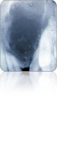
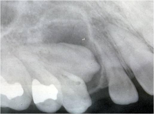
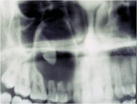
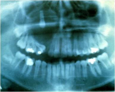
Ill-defined 3 X 3 cm radiolucency in left maxilla in teeth numbers 10 - 11 area
Both teeth test non-vital
Diagnosis
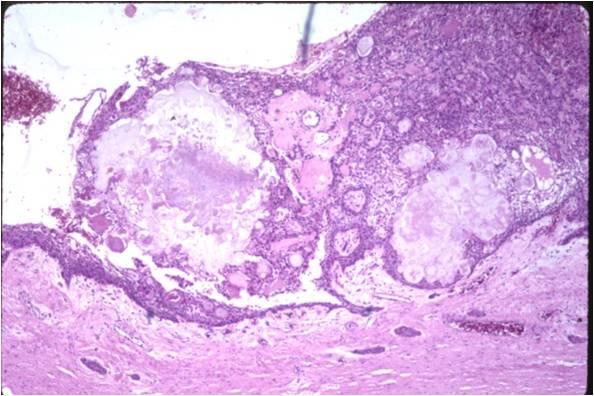
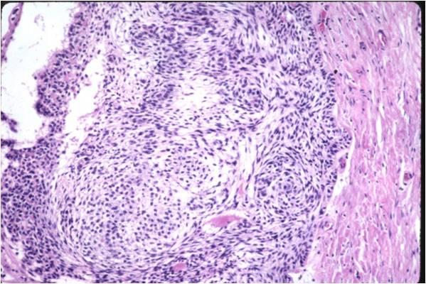
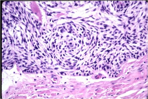
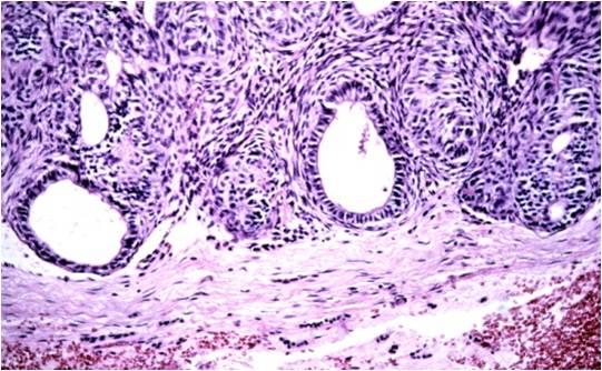
Adenomatoid Odontogenic Tumor (AOT)
Clinical Features
An uncommon benign epithelial odontogenic lesion
First described in 1905
Several synonyms have been used:
Adenoameloblastoma and Odontogenic adenomatoid tumor
Current WHO classification favors AOT
Most common in teens
F:M 2:1; Max:Mand 2:1
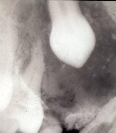
Most often associated with an impacted tooth
A demarcated radiolucency mimicking a dentigerous cyst
Occasional spotty radio-opacities are seen and suggest the dx
Treatment & Prognosis
Usually these are small but some can be large if not observed/treated
Most OMFP do not consider these to be neoplasms but more like a hyperplastic cyst
Histology is characteristic; they are encapsulated; the nature of the calcifications is not known
Treatment is excision
Recurrence is virtually zero
References
•Melrose, R J: Epithelial Odontogenic Tumors. Seminars in Diagnostic Pathology 1999;16:271-287
•Abrams AM, Melrose, RJ, Howell FV: Adenoameloblastoma: A Clinicopathologic study and report of 10 new cases. Cancer; 1968, 22:175-185
•Leon JE, Mata GM, Fregnani ER, et al: Clinicopathologic and immunohistochemical study of 39 cases of adenomatoid odontogenic tumor: a multicentric study, Oral Oncol 41: 835-842, 2005
Courtney RM, Kerr DA The Odontogenic Adenomatoid Tumor} A comprehensive review of 21 cases, Oral Surg Oral Med Oral Pathol 39:424-435, 1975