CPC Case I
Provided By: Satish Kumar DDS, MDSc - 2012-05-16
Clinical History
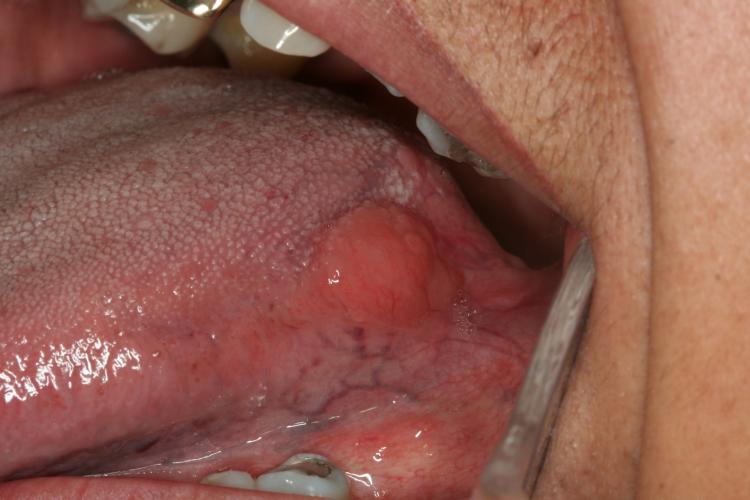
47-year-old Guatemalan female referred by her general dentist for evaluation of an a asymptomatic growth on the left lateral border of the tongue which had been present for 4 months.
Past Medical History: Heart murmur diagnosed in childhood and recent history of neurocysticercosis which was successfully treated.
Clinical Examination: Extraoral within normal limits. Intraoral reveals a single well defined swelling on the left lateral border of the tongue measuring about 1 cm x 0.5 cm, with normal color and smooth surface, soft and non-tender on palpation.
Differential Diagnosis
-
Schwannoma
-
Cysticercosis of the tongue
-
Granular Cell Tumor
-
Solitary Neurofibroma
-
Salivary Gland Neoplasm:
-
Pleomorphic adenoma
-
Mucoepidermoid Carcinoma
Diagnosis
-
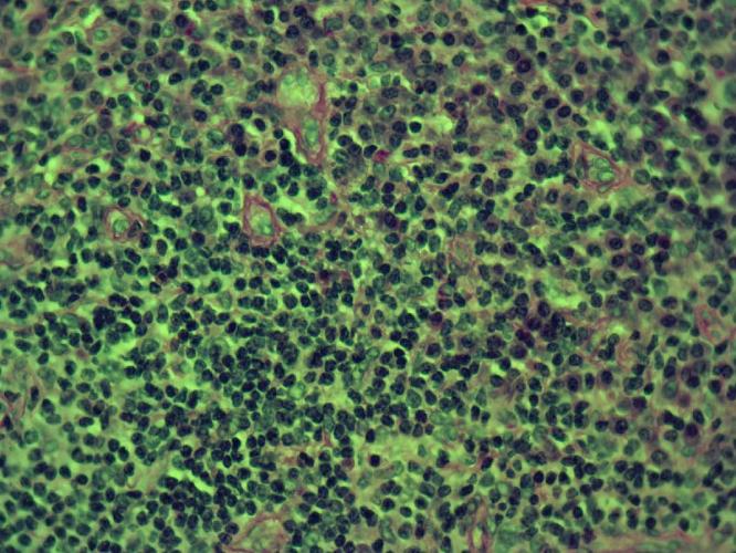
Marginal Zone B�]cell Lymphoma with plasmocytoid features

Clinical Features
Lymphomas: group of malignancies originating in the lymphatic system.
Somatic mutation in a lymphocyte progenitor
The progeny of the affected cell usually carries the phenotype of a B, T, or natural killer cell as determined by immunophenotyping and/or gene rearrangement studies.
Lymphomas are categorized into two main types: Hodgkin�fs Lymphomas, Non Hodgkin Lymphomas (NHL).The later, may initially developed within the lymphatic tissues and subsequently progresses to an extranodal site
Radiographic Findings
Treatment & Prognosis
-
Referred to Norris Cancer Center for furtherevaluation and treatment
-
No other sites of malignant involvement
-
Repeated biopsies confirmed diagnosis
-
Surgical excision and chemotherapy
-
On surveillance and no recurrence
Discussion
Lymphomas in Head & Neck Region
Second most common neoplasia, after squamous cell carcinoma.
~ 3.5% of all oral malignancies and the most common site is the tonsil followed by the parotid gland.
When found in the oral cavity they usually represent a part of an already disseminated disease.
In some cases it represents a localized disease process.
References