39th CPC - Case #1
Provided By: Ameet Chopra DDS - 2009-05-27
Clinical History
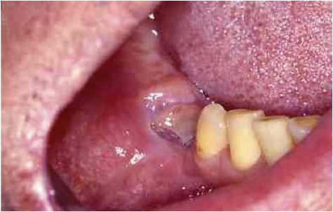
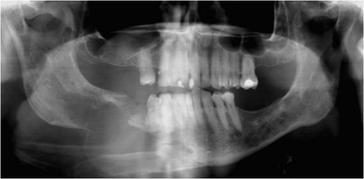
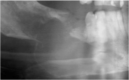
62 year old male presents with the chief complaint that he has a growth in his right lower jaw. His past medical history is unremarkable and he states that the growth has been present for months now but is painless. Review of systems is non-contributory. Intra oral exam reveals a firm swelling of the right mandible that is slightly tender to palpation. The right mandible could be manipulated during palpation in a manner that indicated pathologic fracture, which can also be appreciated on the panoramic radiograph. Extra oral exam reveals asymmetry and expansion in the region of the right mandible and submandibular space, and jaw deviation is observed during mouth opening. No lymphadenopathy is noted.
Unaswered questions
Unaswered questions
- Any pain?
- Any paresthesia?
- Recent tooth extraction?
- In depth review of medical history
- Family history of cancer?

Differential Diagnosis
- Developmental
- Keratocyst (Basal Cell Nevus syndrome)
- Inflammatory
- Osteomyelitis
- Reactive
- Osteonecrosis of jaw
- Hyperparathyroidism
- Neoplasm
- Multiple Myeloma
- Mestatic Carcinoma
- Ostesarcoma
- Intraosseous minor salivary gland tumor
- Lymphoma
- Leukemia
- Multiple Myeloma
- Misc
- Langerhan's cell histiocytosis
- Usually seen as slow growing, well-defined lesions
- Root resorption, tooth mobility, paresthesia
- Pathologic fracture is rare
- Occurs as solitary lesions, although syndromes are present with multiple lesions
- Basal Cell Nevus Syndrome
- Chronic osteomyelitis and chronic osteitis are not prime suspects because there are no manifestations of infection, such as local pain, cervical lymphadenitis, or leukocytosis
- Xray shows lytic center with a ring of sclerosis
- Often seen with mandible fracture
- Osteonecrosis
- Bisphosphonate induced
- Radiation (ORN) Unlikely given no history of previous radiation therapy or treatment with bisphophonates
- Patients generally present with pain
- Can see exposed necrotic bone
- Abnormal proliferation of certain cell types
- Usually rapid growth associated with pain, paresthesia, loose teeth, can see pathologic fracture
- Slow growing indicative of benign process, while rapid usually malignant
- Malignancy of plasma cell origin that often appears to have a multicentric origin with bone (bone marrow). Characterized by proliferation of a single clone of abnormal plasma cells
- Cause is unknown. Accounts for nearly 50% of all malignancies involving bone (excluding metastatic disease)
- Signs and symptoms of this disease result from the uncontrolled proliferation of the tumor cells and manufacture of their protein products
- Disease of adults, with men slightly more affected than women. The median age is between 60 and 70
- Bone pain is the most common symptom. Pathologic fracture of affected bones have been reported
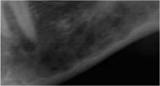
- Radiographs show multiple well-defined, punched-out radiolucencies. Any bone may be affected with the jaws involved in 30% of cases
- Renal failure may be present 2/2 accumulation of proteins (Bence Jones proteins)
- The suspicion of metastatic tumor is enhanced by symptoms of a primary tumor elsewhere in the body or history of previous tumor treatment
- Metastatic bone lesions may assume a variety of radiographic appearances, one of which is multiple, small, well-defined radiolucencies
- Malignant cells from distant sites, usually are carried from their primary site to the jaws via the paravertebral veins of Batson
- Incidence of metastasis to Jaws
- Breast 31%
- Lung 27%
- Kidney 14%
- Bone 10%
- Prostate 5%
- Metastatic Malignancies of Jaws
- Mobile teeth
- Pain
- Radiolucencies
- Radiopacities
- Fracture
- Paresthesia
- aka Langerhan's Cell Histiocytosis
- Involves the Langerhan's cells, which are dendritic cells in the skin and mucosa that have a macrophage-like function
- Three different forms
- Letterer-Siwe: acute disseminated form
- Hand-Schuller-Christian: chronic disseminated form
- Eosinophilic granuloma: chronic localized form
- In the chronic forms of the disease, can see multiple punched-out, well defined radiolucencies
Diagnosis
MULTIPLE MYELOMA